Dizziness and Benign Positional Vertigo |
|||||||
|
What is Benign Paroxysmal Positional Vertigo (BPPV)? Benign paroxysmal positional vertigo (BPPV) is a common inner ear disorder that causes brief episodes of vertigo, or spinning sensation. The name BPPV comes from the following:
Symptoms of BPPV The most common symptom of BPPV is a sudden sensation of spinning or whirling. The spinning sensation usually lasts for a few seconds to a few minutes, and it can be accompanied by nausea and vomiting. The episodes of vertigo are often triggered by specific head movements, such as:
Causes of BPPV BPPV is caused by a problem with the tiny crystals (called otoconia) in the inner ear. These crystals normally help the inner ear detect changes in head position. However, if the crystals become dislodged and move into the wrong part of the inner ear, they can send false signals to the brain about the body's balance. This can lead to vertigo. Treatment for BPPV There are a number of treatments available for BPPV. The most common treatment is called the Epley maneuver. The Epley maneuver is a series of head movements that can help to move the crystals back into the correct position in the inner ear. Other treatments for BPPV include the Brandt-Daroff exercises and medication. Outlook for BPPV In most cases, BPPV can be treated successfully. However, it is possible for the condition to come back. If you experience symptoms of BPPV, it is important to see a doctor to get a diagnosis and treatment. The inner ear and how it works The inner ear is responsible for balance and hearing. It is made up of three parts:
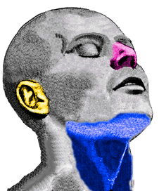
The external ear consists of the auricle (also known as the pinna), which is the visible part of the ear, and the external auditory canal, which is a tube that leads to the tympanic membrane (eardrum). The middle ear includes the tympanic membrane and the three ossicles, which are the malleus ("hammer"), incus ("anvil"), and stapes ("stirrup"). These bones transmit sound vibrations from the tympanic membrane to the inner ear.
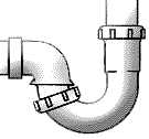
The inner ear is a fluid-filled series of chambers. One of these chambers, the cochlea, is responsible for converting sound vibrations into nerve impulses. The brain interprets these nerve impulses as sound and what we call "hearing." The inner ear also contains three semicircular canals which are responsible, in part, for sensing movement and maintaining balance. As you can see, these three canals (named anterior, lateral, and posterior) are oriented at roughly right angles to one another. The movement of the fluid within these canals allows the brain to sense rotation of the head through all three directions in space (e.g., left-right, forward-back, and up-down). All three canals are connected to a large chamber called the vestibule. It has been discovered that the probable cause of BPPV is the dislodgement of small calcium carbonate crystals that float through the inner ear fluid and strike against sensitive nerve endings (the cupula) within the balance apparatus at the end of each semicircular canal (the ampulla). (Another name for BPPV is cupulolithiasis, meaning "rocks in the cupula"). These crystals, known as otoconia, usually dissolve or fall back into the vestibule within several weeks and no longer cause any symptoms. However, in some patients, these crystals become trapped in the fluid of the balance chamber and periodically cause symptoms, as gravity and head movements cause them to repeatedly strike against the cupula. In these patients, the symptoms may not subside and they become severely incapacitated. Interestingly, the loose otoconia tend to settle preferentially within the posterior semicircular canal. As you can imagine from looking at the illustration above, this is because the posterior canal hangs down like the water trap in a drain pipe, allowing the crystals to settle in the bottom of the canal.
How is BPPV diagnosed? Benign paroxysmal positional vertigo (BPPV) is a common inner ear disorder that causes sudden episodes of vertigo, or dizziness when you change your head position. The most important means for diagnosing BPPV is the physical examination and history of the patient. A patient with dizziness or vertigo without hearing problems suggests the diagnosis of BPPV. A normal ear exam, audiogram, and neurological exam are expected. A simple positional test, performed in the doctor's office, is usually all that is needed to confirm the diagnosis of BPPV. One such test is the Dix-Hallpike test. In this test, the patient is seated upright on an examination table with their head turned 45 degrees to one side. The examiner then quickly lowers the patient's head back until it is hanging over the edge of the table. If the patient has BPPV, they will experience a characteristic movement of the eyes, called nystagmus, within a few seconds. The nystagmus will typically last for 15 to 20 seconds. If the nystagmus is seen and the patient becomes dizzy, then the ear which is pointing toward the floor is the one with the loose otoconia. If no nystagmus is seen, the examiner will repeat the test, this time turning the head to the opposite side. Occasionally, in order to confirm the extent of the inner ear dysfunction, an electronystagmogram (ENG) will be ordered. An ENG is a test that records the eye movements of a patient while they are in different head positions. This test can help to confirm the diagnosis of BPPV and identify the specific ear that is affected. What are the treatments for BPPV? Once tests have confirmed the diagnosis of BPPV and the affected ear, patients are instructed to avoid lying down on the affected side. Usually, medications like Antivert (meclizine), Dramamine, Valium, or Phenergan are not recommended because they cause sedation. By carefully avoiding the provocative position, patients can usually avoid bringing about the symptoms. If left untreated, the condition usually clears within several weeks. The Epley or Semont Maneuver Recently, researchers have found that a simple and well-tolerated physical therapy technique performed in the office can relieve vertigo in a high percentage of patients. The Otolith Repositioning Procedure of Semont and Epley has become well accepted and is based on using gravity to move the crystals away from the nerve endings into an area of the inner ear that will not cause vertigo. There are several variations of this manoeuvre, but they all involve moving the patient's head in a specific way to reposition the otoconia. If you are experiencing dizziness or vertigo, it is important to see a doctor to get a diagnosis and treatment. BPPV is a treatable condition, and most people experience relief from symptoms after a few weeks. |
|||||||